Autophagy: Can Fasting Help the Brain Clean Up?
Cells break down and reuse damaged parts through a process called autophagy. Fasting can stimulate it in mice; whether an overnight fast helps the human brain remains an open question.
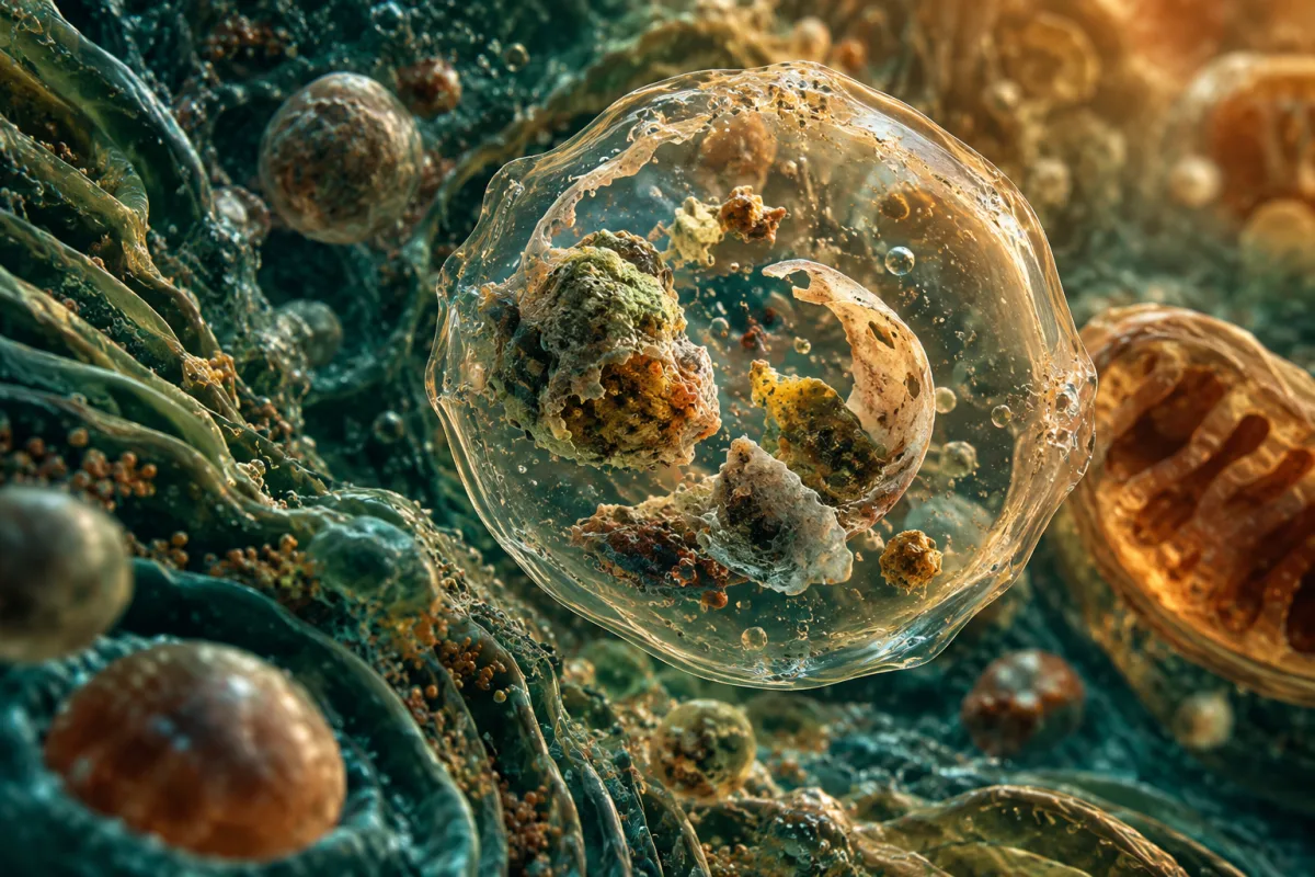
Finish dinner early, skip the late-night snack and give your brain time to clean itself. It is an appealing argument for fasting, especially when it comes with the promise of protection against Alzheimer’s disease. Behind it lies a real biological process called autophagy: the way cells break down and reuse their own damaged parts.
The difficulty is knowing how far the evidence takes us. Researchers have seen fasting stimulate this process in mouse neurons. Small human studies have detected changes in blood and muscle that may help explain how our bodies respond to going without food. They have not established a fasting schedule that clears harmful proteins from the human brain.
That distinction can disappear in accounts that describe autophagy as a switch, turning on after 12 hours and reaching its peak a few hours later. The research tells a more interesting, and less certain, story.
The recycling that never stops
Inside a cell, worn-out components are enclosed in a small membrane-bound compartment called an autophagosome. This delivers them to a lysosome, where enzymes break them down. The resulting materials can be used again.
Scientists call this particular form of recycling macroautophagy, usually shortened to autophagy. Its importance was recognised with the 2016 Nobel Prize in Physiology or Medicine, awarded to Yoshinori Ohsumi for work identifying the genes behind the process in yeast. Much of the same machinery operates in mammalian cells.
Autophagy carries on even when food is plentiful. Cells can increase it when nutrients or energy become scarce, regulated by systems that sense their condition. Among these are mTOR, which tends to restrain autophagy when nutrients and growth signals are abundant, and AMPK, which responds to low energy. A 2020 review in the New England Journal of Medicine describes how starvation and aerobic exercise can increase the process.
For neurons, which generally have to last a lifetime, this ongoing maintenance is particularly important. Two mouse studies published in Nature in 2006 showed what happened when researchers disabled essential autophagy genes in the nervous system: abnormal proteins accumulated, movement deteriorated and neurons were lost (Hara et al.; Komatsu et al.).
The experiments established that neurons need autophagy to stay healthy. Whether increasing it through fasting can prevent disease in people is a separate question.
What happened when mice went without food
One influential experiment came from Alirezaei and colleagues in 2010. They studied mice with a fluorescent marker that allowed them to see autophagosomes inside individual neurons, rather than relying on measurements from the brain as a whole.
After 24 hours without food, autophagosomes were more abundant in cortical neurons. After 48 hours, their numbers in cerebellar Purkinje cells were roughly three to four times those seen in fed mice. Other measurements supported the researchers’ interpretation that fasting had stimulated autophagy (Alirezaei et al., 2010).
This was an important finding, but it came from small groups of mice subjected to an acute fast. A day without food places a substantial metabolic demand on a mouse. There is no simple conversion that turns those results into a 12-hour recommendation for a person.
The measurements also require care. Counting autophagosomes reveals how many recycling compartments are present at a particular moment. More compartments can mean that cells are making them faster, or that they are clearing them more slowly. The additional measurements favoured increased production in this experiment, but the study did not directly quantify how much damaged protein had been destroyed.
It did not establish an hourly timetable for different brain regions, either. Claims of a 300 percent increase in human brain autophagy after exactly 12 hours go beyond what these mice can tell us.
The human clues are still indirect
Measuring the same process in people is much harder. A blood sample cannot reveal the rate at which a living brain is recycling material. A 2022 review of acute fasting concluded that a single fast can alter glucose and fat metabolism within 24 hours, while its ability to meaningfully increase autophagy remained uncertain.
A small trial published in 2019 offers one of the more frequently cited human findings. Eleven adults with overweight tried two eating schedules for four days each. In one, they ate between 8 a.m. and 2 p.m.; in the other, between 8 a.m. and 8 p.m. The food was matched, allowing researchers to examine meal timing rather than a difference in intake.
After the longer overnight fast, morning blood samples showed about 22 percent higher expression of LC3A, a gene involved in autophagy. The finding was exploratory, and the authors cautioned that gene expression does not necessarily reflect protein levels or activity of the process itself. They had measured blood cells, not brain cells (Jamshed et al., 2019).
Other tissues have produced different results. A 2022 study found increased autophagy markers in the livers of fasting mice, but not in their muscles. In its human arm, involving 50 women following intermittent-fasting schedules for eight weeks, muscle samples did not show a clear overall activation of the markers studied; some were lower (Chaudhary et al., 2022).
These studies help researchers work out where and how fasting acts. They also show why a result from one tissue cannot be assumed to apply throughout the body.
Evidence for sharper thinking is similarly unsettled. A 2024 systematic review examined human studies of calorie restriction and intermittent fasting that measured BDNF, a protein involved in neuronal function. Of 16 studies with usable data, five reported an increase, five a decrease and six no clear change. Cognitive results were also mixed. Feeling more alert before breakfast may be someone’s experience, but it does not establish that autophagy has improved their brain function.
Why sleep enters the conversation
Discussions of fasting often bring in another form of brain waste clearance: the glymphatic system. Unlike autophagy, which operates inside cells, this involves fluid moving through brain tissue and carrying away soluble material between cells.
In a widely cited 2013 mouse study, sleep or anaesthesia was associated with an expansion of the space between cells and faster clearance of beta-amyloid, a protein implicated in Alzheimer’s disease. Human studies have offered clues too, although the pathway is difficult to observe directly (Benveniste et al., 2018).
One experiment compared brain scans from 20 healthy adults after a rested night and after a night without sleep. The sleepless night was followed by an average increase of about 5 percent in the amyloid imaging signal in a region covering the right hippocampus and thalamus. Responses varied considerably. This was a short-term change in a tracer signal, rather than a measurement of new plaques or future dementia risk (Shokri-Kojori et al., 2018).
Autophagy
Cells package and break down their own components. Fasting experiments in mouse neurons show increases in the compartments involved in this process.
Glymphatic clearance
Fluid movement through brain tissue helps carry away soluble waste. Sleep studies investigate this pathway separately from autophagy.
Together, these findings give researchers reasons to investigate sleep and brain health. They do not show that fasting overnight adds a second, measurable dose of brain cleaning, or that a late snack shuts autophagy down for six to eight hours. The sleep duration article looks more closely at the human evidence on sleep and longevity.
A metabolic change is not proof of brain protection
Going without food does change the body’s fuel supply. As liver glycogen falls, fat use increases and the liver produces ketones, which the brain can use for energy. A 2019 review of intermittent fasting describes this metabolic shift alongside cellular responses observed in animal research.
Its timing varies with the previous meal, activity, glycogen stores and metabolic health. In the eleven-person meal-timing trial, the longer fast increased morning ketones by about 0.03 millimoles per litre. That was a detectable change, but the trial did not test whether it improved memory or reduced Alzheimer’s pathology.
The distinction matters when fasting is promoted as disease prevention. None of the studies discussed here establishes that regularly fasting for 12 to 24 hours reduces amyloid and tau accumulation by 40 percent, or halves brain inflammation within a day.
What this means for everyday eating
A roughly 12-hour gap between dinner and breakfast already fits many people’s routines. There is no established brain-autophagy threshold that everyone needs to reach, and no evidence here that extending that gap will prevent Alzheimer’s disease.
Some people find a shorter eating window useful for managing evening snacking or overall intake. Those are reasonable outcomes to assess in their own right. The caloric-restriction article explains why meal timing, calorie intake and nutritional quality need to be considered separately.
Children, people who are pregnant or breastfeeding, those who are underweight or frail, and anyone with a current or past eating disorder should avoid experimenting with prolonged fasting. People taking insulin or other medicines that can cause low blood glucose need individual medical advice before changing their eating schedule.
The research also offers no basis for treating supplements as proven ways to increase autophagy for better brain health. A compound’s effect on a pathway in a laboratory does not establish a useful dose or a health benefit in people. The supplement review examines that gap.
Autophagy remains a promising area of research because it addresses a fundamental problem of ageing: how cells maintain themselves over time. The mouse experiments make fasting worth studying. The human evidence has yet to turn that possibility into a reliable prescription.
For now, the science gives us a reason to be curious about fasting, but not a reason to watch the clock for the moment our brains supposedly start to clean themselves.
Editorial note: This article was revised with AI-assisted editing.
Sources
- Nobel Prize in Physiology or Medicine 2016: mechanisms of autophagy
- Mizushima and Levine, 2020: Autophagy in human diseases
- Hara et al., 2006: Loss of basal autophagy in neural cells causes neurodegeneration in mice
- Komatsu et al., 2006: Loss of autophagy in the central nervous system causes neurodegeneration in mice
- Alirezaei et al., 2010: Short-term fasting induces profound neuronal autophagy
- Stratton et al., 2022: Physiological responses to acute fasting
- Jamshed et al., 2019: Early time-restricted feeding and markers of autophagy in humans
- Chaudhary et al., 2022: Intermittent fasting and autophagy markers in mouse liver and in mouse and human muscle
- Alkurd et al., 2024: Calorie restriction, intermittent fasting, BDNF, and cognition
- de Cabo and Mattson, 2019: Effects of intermittent fasting on health, aging, and disease
- Xie et al., 2013: Sleep drives metabolite clearance from the adult brain
- Shokri-Kojori et al., 2018: β-Amyloid accumulation after one night of sleep deprivation
- Benveniste et al., 2018: The glymphatic system and waste clearance with brain aging
- Longtime: caloric restriction and the human evidence
- Longtime: sleep duration and longevity
- Longtime: supplements for longevity